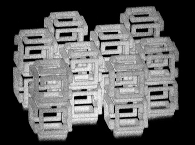
Using a screwdriver on the tiny microcircuits arrayed inside a computer hard drive can be a real eye strain. Even more challenging is building the microcircuits or other electronic components at the nanoscale, one-billionth of a meter or less. That’s why researchers are always on the lookout for new tools to help them work on such a minute scale. But some of these incredibly tiny tools and scaffolds can derive from very unexpected sources.

As published in the journal Science, an NIH-funded team has developed a technique called implosion fabrication to build impressively small and intricate components on the nanoscale [1]. Its secret ingredient: water-swollen gels that you’d find in a baby’s disposable diaper. A baby’s disposable diaper? If that sounds familiar, my blog highlighted a related technique called expansion microscopy a few years ago that uses water-swollen gels that are generated from a compound used in diapers called sodium polyacrylate.
The previously-reported microscopy technique, from the lab of Edward Boyden, Massachusetts Institute of Technology, Cambridge, embeds biological samples in a fine web of sodium polyacrylate. When water is added, the gel expands, blowing up the specimen to 100 times its original size. This groundbreaking technique, called expansion microscopy, has enabled labs around the world to use conventional microscopes for high-resolution, nanoscale imaging.
In the latest work, Boyden’s team, including co-first authors Daniel Oran and Samuel Rodriques, asked a simple question: What would happen if they applied the sample preparation technique used for expansion microscopy—only in reverse? To find out, Boyden’s team created millimeter-sized blocks of the super-absorbent sodium polyacrylate diaper compound. After using a nifty trick for attaching molecular anchors in a 3D pattern, they dehydrated the gel and voila! The structures imploded and shrank down to one-thousandth their original size, while holding their 3D shape. During the process, they can add to the anchors a range of functional molecules or elements. These include DNA, nanoparticles, semiconductors, or almost anything that’s needed.
While more work is needed to perfect the new technique, the researchers have already shown it can create objects one cubic millimeter in size, engineered to include intricate details down to about 50 nanometers.For comparison, a virus is about 30 to 50 nanometers.
These latest findings come as a reminder that advances in biomedicine often lead in wonderful and unexpected new directions. Out of the NIH-funded efforts related to The Brain Research through Advancing Innovative Neurotechnologies® (BRAIN) Initiative, members of the Boyden Lab wanted to see the brain better using basic microscopes. Now, we have a widely-applicable promising new approach to nanofabrication.
[1] 3D nanofabrication by volumetric deposition and controlled shrinkage of patterned scaffolds. Oran D, Rodriques SG, Gao R, Asano S, Skylar-Scott MA, Chen F, Tillberg PW, Marblestone AH, Boyden ES. Science. 2018 Dec 14;362(6420):1281-1285.
