An official website of the United States government
Here’s how you know
Official websites use .gov A
.gov
website belongs to an official government organization in the United States.
Secure .gov websites use HTTPS A
lock
(
) or
https://
means you’ve safely connected to the .gov website. Share sensitive information only on official, secure websites.
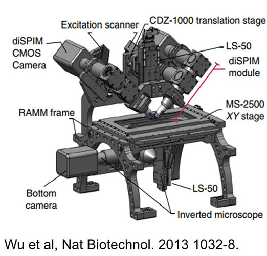
The DISPIM is a dual light sheet microscope that acquires two orthogonal views of a sample. These two views can then be fused and joint deconvolved to generate a single volume with high isotropic resolution. Like other light sheet microscopes, the DISPIM provides rapid volume imaging with a low photodose and is therefore ideally suited for long term or high repetition rate imaging in samples where phototoxicity/photobleaching can be a problem. Unlike conventional light sheet microscopes, the joint deconvolution process provides good resolution in all directions.
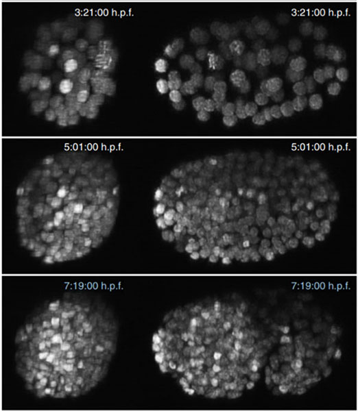
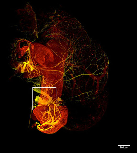
Image Gallery
Figure 1. Spatial resolution: 330nm isotropic. Temporal resolution: 200Hz framerate, 1-5Hz volumetric. Excitation wavelengths: 405nm, 488nm, 561nm, 639nm. DISPIM is ideal for imaging experiments in which photobleaching or phototoxicity is a critical issue. This is the fastest microscope currently available at AIM.
Figure 2. 40X 0.8NA, dual views provide 330nm isotropic resolution with low photobleaching/phototoxicity. Ideal for prolonged imaging of live samples.
3. 17X 0.4NA, dual views provide lower resolution (~1um lateral) with a larger FOV (800x800um). Image plane can be scanned rapidly through a large sample of cleared tissue (up to 1cm x 1cm x 5mm). Refractive index: 1.33-1.56. Cleared mouse heart (courtesy Will Kowalski, NHLBI). 1.6mm x 2.6mm x 780um. Yellow: Anti-Tuj1-Alexa488 (pan-neuronal), red: Anti-SM22alpha-Alexa594 (smooth muscle) Acquisition time: 608s for 3 colors. iDISCO.